Sunday, 14 June 2015

Esther Gokhale's Five Tips For Better Posture And Less Back Pain

Esther Gokhale's Five Tips For Better Posture And Less Back Pain

From NPR June 14/2015
http://www.npr.org/sections/goatsandsoda/2015/06/08/412314701/lost-posture-why-indigenous-cultures-dont-have-back-pain?utm_source=npr_newsletter&utm_medium=email&utm_content=20150614&utm_campaign=mostemailed&utm_term=nprnews


Try these exercises while you're working at your desk, sitting at the dinner table or walking around, Esther Gokhale recommends.

1. Do a shoulder roll: Americans tend to scrunch their shoulders forward, so our arms are in front of our bodies. That's not how people in indigenous cultures carry their arms, Gokhale says. To fix that, gently pull your shoulders up, push them back and then let them drop — like a shoulder roll. Now your arms should dangle by your side, with your thumbs pointing out. "This is the way all your ancestors parked their shoulders," she says. "This is the natural architecture for our species."

2. Lengthen your spine: Adding extra length to your spine is easy, Gokhale says. Being careful not to arch your back, take a deep breath in and grow tall. Then maintain that height as you exhale. Repeat: Breathe in, grow even taller and maintain that new height as you exhale. "It takes some effort, but it really strengthens your abdominal muscles," Gokhale says.

3. Squeeze, squeeze your glute muscles when you walk: In many indigenous cultures, people squeeze their gluteus medius muscles every time they take a step. That's one reason they have such shapely buttocks muscles that support their lower backs. Gokhale says you can start developing the same type of derrière by tightening the buttocks muscles when you take each step. "The gluteus medius is the one you're after here. It's the one high up on your bum," Gokhale says. "It's the muscle that keeps you perky, at any age."

4. Don't put your chin up: Instead, add length to your neck by taking a lightweight object, like a bean bag or folded washcloth, and balance it on the top of your crown. Try to push your head against the object. "This will lengthen the back of your neck and allow your chin to angle down — not in an exaggerated way, but in a relaxed manner," Gokhale says.

5. Don't sit up straight! "That's just arching your back and getting you into all sorts of trouble," Gokhale says. Instead do a shoulder roll to open up the chest and take a deep breath to stretch and lengthen the spine.

[Added June 10] 
So Gokhale studied findings from anthropologists, such as Noelle Perez-Christiaens, who analyzed postures of indigenous populations. And she studied physiotherapy methods, such as the Alexander Technique and the Feldenkrais Method.

And the original post  ...

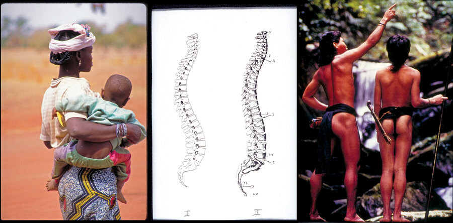
Primal posture: 

 Ubong tribesmen in Borneo (right) display the perfect J-shaped spines. 
A woman in Burkina Faso (left) holds her baby so that his spine stays straight. The center image shows the S-shaped spine drawn in a modern anatomy book (Fig. I) and the J-shaped spine (Fig. II) drawn in the 1897 anatomy book Traite d'Anatomie Humaine.
Courtesy of Esther Gokhale and Ian Mackenzie/Nomads of the Dawn 
 Primal posture: Ubong tribesmen in Borneo (right) display the perfect J-shaped spines. A woman in Burkina Faso (left) holds her baby so that his spine stays straight. The center image shows the S-shaped spine drawn in a modern anatomy book (Fig. I) and the J-shaped spine (Fig. II) drawn in the 1897 anatomy book Traite d'Anatomie Humaine.
 
 
Editor's note, June 10: We have added an acknowledgement of several sources that Esther Gokhale used while developing her theories on back pain. These include physiotherapy methods, such as the Alexander Technique and the Feldenkrais Method, and the work of anthropologist Noelle Perez-Christiaens.
Back pain is a tricky beast. Most Americans will at some point have a problem with their backs. And for an unlucky third, treatments won't work, and the problem will become chronic.
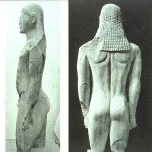
Many ancient statues, such as this one from Greece, display a J-shaped spine. The statue's back is nearly flat until the bottom, where it curves so the buttocks are behind the spine. i
Many ancient statues, such as this one from Greece, display a J-shaped spine. The statue's back is nearly flat until the bottom, where it curves so the buttocks are behind the spine.
Courtesy of Esther Gokhale/Gerard Mackworth-Young 
 
Believe it or not, there are a few cultures in the world where back pain hardly exists. One indigenous tribe in central India reported essentially none. And the discs in their backs showed little signs of degeneration as people aged.

An acupuncturist in Palo Alto, Calif., thinks she has figured out why. She has traveled around the world studying cultures with low rates of back pain — how they stand, sit and walk. Now she's sharing their secrets with back pain sufferers across the U.S.

About two decades ago, Esther Gokhale started to struggle with her own back after she had her first child. "I had excruciating pain. I couldn't sleep at night," she says. "I was walking around the block every two hours. I was just crippled."

Gokhale had a herniated disc. Eventually she had surgery to fix it. But a year later, it happened again. "They wanted to do another back surgery. You don't want to make a habit out of back surgery," she says.

This time around, Gokhale wanted to find a permanent fix for her back. And she wasn't convinced Western medicine could do that. So Gokhale started to think outside the box.

She had an idea: "Go to populations where they don't have these huge problems and see what they're doing."


Then over the next decade, Gokhale went to cultures around the world that live far away from modern life. She went to the mountains in Ecuador, tiny fishing towns in Portugal and remote villages of West Africa.

"I went to villages where every kid under age 4 was crying because they were frightened to see somebody with white skin — they'd never seen a white person before," she says.

Gokhale took photos and videos of people who walked with water buckets on their heads, collected firewood or sat on the ground weaving, for hours.

"I have a picture in my book of these two women who spend seven to nine hours everyday, bent over, gathering water chestnuts," Gokhale says. "They're quite old. But the truth is they don't have a back pain."

She tried to figure out what all these different people had in common. The first thing that popped out was the shape of their spines. "They have this regal posture, and it's very compelling."

And it's quite different than American spines.

If you look at an American's spine from the side, or profile, it's shaped like the letter S. It curves at the top and then back again at the bottom.

But Gokhale didn't see those two big curves in people who don't have back pain. "That S shape is actually not natural," she says. "It's a J-shaped spine that you want."

In fact, if you look at drawings from Leonardo da Vinci — or a Gray's Anatomy book from 1901 — the spine isn't shaped like a sharp, curvy S. It's much flatter, all the way down the back. Then at the bottom, it curves to stick the buttocks out. So the spine looks more like the letter J.

"The J-shaped spine is what you see in Greek statues. It's what you see in young children. It's good design," Gokhale says.

So Gokhale worked to get her spine into the J shape. And gradually her back pain went away.


Healthy spines in the Western world: The J-shaped spine is often seen in photographs from the late 19th and early 20th centuries. i
Healthy spines in the Western world: The J-shaped spine is often seen in photographs from the late 19th and early 20th centuries.

Wednesday, 6 May 2015

temporary Links to cognitive articles


 Look Them up

The Medical Biochemistry Page

Brain-derived neurotrophic factor - Wikipedia, the free encyclopedia

Selank dosage, side effects and where to buy

PEPTIDE PROFILES - Evolutionary.org

Sermorelin Dosage and Side Effects

Nootropics – Selank Peptide Explained | IronMag™ - Bodybuilding Science, Fitness, Sports, and Anabolic Steroid Resource


Bremelanotide PT-141 Guide | Dosage Information

Updated Selank Trial | Metabolic Alchemy

MT2, Melanotan II (Afamelanotide)

Trade Names:
MT2, Melanotan
Chemical Names:
Afamelanotide
Routes:
Subcutaneous Injection
Melanotan II (MT2) was first synthesized at the University of Arizona in the 1980’s. Melanotan II is a peptide originally developed as a skin tanning agent, but subsequently investigated as a potential treatment for sexual dysfunction. 

To date there is no medically approved drug that contains the Melanotan II peptide, instead all available products are from commercial labs that are allowed to produce and distribute peptides as research products.

Even though the only Melanotan II peptides  available to the general public are unlicensed and unregulated drugs, thousands of people use it daily. A number of health and drug control agencies around the world issued warnings to the potential of health risks associated with unregulated medications.
Regardless of all the warnings the popularity of MT2 seems to be on the increase. One obvious reason for the popularity of MT2 is that it actually works and results are noticed fairly quickly. 
Another cause for the thriving trade of MT2 is that it’s not illegal to buy, import or use in most countries.

Since there’s no brand name  product available no official guideline has yet been established on the correct usage of this peptide. However through trial and error and experimentation the MT2 community developed a variety of protocols. The most popular and effective protocols are those that make use of a “loading” phase followed by a “maintenance” phase.

The loading phase is normally limited to about 3 to 5 days and in this time dosages of 0.5 to 1mg are injected subcutaneously nightly. Near the end of this phase one or two tanning sessions are taken so that the tanning process can be kick started. It’s very important to expose all parts of the body to equal amounts of sunlight or ultraviolet radiation. 

This is to allow for a uniform tan and to limit chances of dark or lighter spots. Results are rapid and care should be taken not to overdo usage  as that will result in an abnormally dark appearance.

The purpose of the maintenance phase is to prolong the period for which the tanned skin is required. Usually a dose of 0.5mg is taken 1-2 times per week. This might be altered depending on how the person reacts. If the tan is getting lighter the dosage might be increased or the period between injections shortened. If the tan is getting to dark the dosage can be lowered or the injection frequency decreased.


A number of initial side-effects can be expected when first using MT2, this is due to the structure of the molecule not entirely mimicking that of natural human a-MSH (alpha-melanocyte stimulating hormone) hormone.

 The body’s immune system will recognize it as being foreign and thus an allergic response may occur.

Facial flushing similar to initial Vitamin B3 Niacin non flushing formula. 
The melanotan flushing effects are less pronounced and usually transient within 30-45 minutes. Again prophylactic use of a small dose of benedryll  prior to using the peptide will eliminate or lower this initial side possible side effect.
Allerdryl/benedryl  can be  used as a preventive for those people who are especially sensitive to taking MT2.
Just as well, much lower concentrations of the injected drug would also lessen the side-effects.

 Some people find that taking the first few shots at night before bed lowers or reduces the initial flushing side effects.
Any transient nausea can be eliminated by ingesting a small amount of ginger, prior to using the peptide.

 The lyophilized powder may be stored at 4 C for several months . When you reconstitute to nominal volume and store at -20 C, the reconstituted product should be stable for 24 months. 
Best long-term storage solution is to store in lyophilized powder form at -20C. The reconstituted solution is best injected with an insulin syringe.

Use a 1 Ml insulin with a 31 gauge needle. 
Start with 0.5 Iu to 1.0 Iu on the scale of 10.

Reconstituting with 2.5 ml of sterile Na solution will allow for proper dosing.
5-10Iu on a regular 100Iu insulin syringe usually works best.

As with any herbal, peptide or medication always go slow to gauge individual sensitivity.

If you feel unwell, always seek medical attention, call the Telehealth 24/7 

1-866-797-0000     TTY : 1-866-797-0007


Gut Feelings–the "Second Brain" in Our Gastrointestinal Systems [Excerpt]

Gut Feelings–the "Second Brain" in Our Gastrointestinal Systems [Excerpt]

There is a superhighway between the brain and GI system that holds great sway over humans


Penguin Press

More on this Topic

From The Good Gut: Taking Control of Your Weight, Your Mood and Your Long-Term Health, by Justin Sonnenburg and Erica Sonnenburg, PhDs. Reprinted by arrangement with Penguin Press, a member of Penguin Group (USA), LLC, a Penguin Random House Company. Copyright © Justin Sonnenburg and Erica Sonnenburg, 2015.


A primal connection exists between our brain and our gut. 


We often talk about a “gut feeling” when we meet someone for the first time. 
We’re told to “trust our gut instinct” when making a difficult decision or that it’s “gut check time” when faced with a situation that tests our nerve and determination.
This mind-gut connection is not just metaphorical.

 Our brain and gut are connected by an extensive network of neurons and a highway of chemicals and hormones that constantly provide feedback about how hungry we are, whether or not we’re experiencing stress, or if we’ve ingested a disease-causing microbe. 

This information superhighway is called the brain-gut axis and it provides constant updates on the state of affairs at your two ends. That sinking feeling in the pit of your stomach after looking at your postholiday credit card bill is a vivid example of the brain-gut connection at work. 
You’re stressed and your gut knows it—immediately.

The enteric nervous system is often referred to as our body’s second brain. 
There are hundreds of million of neurons connecting the brain to the enteric nervous system, the part of the nervous system that is tasked with controlling the gastrointestinal system. 

This vast web of connections monitors the entire digestive tract from the esophagus to the anus. The enteric nervous system is so extensive that it can operate as an independent entity without input from our central nervous system, although they are in regular communication. 

While our “second” brain cannot compose a symphony or paint a masterpiece the way the brain in our skull can, it does perform an important role in managing the workings of our inner tube. The network of neurons in the gut is as plentiful and complex as the network of neurons in our spinal cord, which may seem overly complex just to keep track of digestion. Why is our gut the only organ in our body that needs its own “brain”? Is it just to manage the process of digestion? Or could it be that one job of our second brain is to listen in on the trillions of microbes residing in the gut?

Operations of the enteric nervous system are overseen by the brain and central nervous system. The central nervous system is in communication with the gut via the sympathetic and parasympathetic branches of the autonomic nervous system, the involuntary arm of the nervous system that controls heart rate, breathing, and digestion. The autonomic nervous system is tasked with the job of regulating the speed at which food transits through the gut, the secretion of acid in our stomach, and the production of mucus on the intestinal lining.

 The hypothalamic-pituitary-adrenal axis, or HPA axis, is another mechanism by which the brain can communicate with the gut to help control digestion through the action of hormones.

This circuitry of neurons, hormones, and chemical neurotransmitters not only sends messages to the brain about the status of our gut, it allows for the brain to directly impact the gut environment. The rate at which food is being moved and how much mucus is lining the gut—both of which can be controlled by the central nervous system—have a direct impact on the environmental conditions the microbiota experiences.

Like any ecosystem inhabited by competing species, the environment within the gut dictates which inhabitants thrive. Just as creatures adapted to a moist rain forest would struggle in the desert, microbes relying on the mucus layer will struggle in a gut where mucus is exceedingly sparse and thin. Bulk up the mucus, and the mucus-adapted microbes can stage a comeback. 
The nervous system, through its ability to affect gut transit time and mucus secretion, can help dictate which microbes inhabit the gut. In this case, even if the decisions are not conscious, it’s mind over microbes.

What about the microbial side? When the microbiota adjusts to a change in diet or to a stress-induced decrease in gut transit time, is the brain made aware of this modification? 

Does the brain-gut axis run in one direction only, with all signals going from brain to gut, or are some signals going the other way? 
Is that voice in your head that is asking for a snack coming from your mind or is it emanating from the insatiable masses in your bowels?

Recent evidence indicates that not only is our brain “aware” of our gut microbes, but these bacteria can influence our perception of the world and alter our behavior. It is becoming clear that the influence of our microbiota reaches far beyond the gut to affect an aspect of our biology few would have predicted—our mind.

For example, the gut microbiota influences the body’s level of the potent neurotransmitter serotonin, which regulates feelings of happiness. Some of the most prescribed drugs in the U.S. for treating anxiety and depression, like Prozac, Zoloft, and Paxil, work by modulating levels of serotonin. And serotonin is likely just one of a numerous biochemical messengers dictating our mood and behavior that the microbiota impacts.

Thursday, 16 April 2015

Delta sleep-inducing peptide


Skip Navigation LinksHome > July 2001 - Volume 18 - Issue 7 > Delta sleep-inducing peptide

European Journal of Anaesthesiology:

Delta sleep-inducing peptide

Pollard, B. J.1; Pomfrett, C. J. D.2

Free Access
Article Outline
Collapse Box

Author Information

1Professor of Anaesthesia, University Department of Anaesthesia, Manchester Royal Infirmary, Oxford Road, Manchester M13 9WL, UK

2Lecturer in the Neurophysiology of Anaesthesia, University Department of Anaesthesia, Manchester Royal Infirmary, Oxford Road, Manchester M13 9WL, UK


Delta sleep-inducing peptide (DSIP) is a naturally occurring substance, which was originally isolated from rabbit brain in 1977 [1]. This curious substance is a nonapeptide that is normally synthesized in the hypothalamus and targets multiple sites including some within the brainstem [2]. 
As its name suggests DSIP promotes sleep and this has been demonstrated in rabbits, mice, rats, cats and human beings [3–5]. In fact DSIP promotes a particular type of sleep which is characterized by an increase in the delta rhythm of the EEG.

DSIP is normally present in minute amounts in the blood. Brain and plasma DSIP concentrations exhibit a marked diurnal variation [6] and there has been shown to be a correlation between DSIP plasma concentrations and circadian rhythm in human beings. Concentrations are low in the mornings and higher in the afternoons. An elevation of endogenous DSIP concentration has been shown to be associated with suppression of both slow-wave sleep and rapid-eye-movement sleep and interestingly also with body temperature [7]. Plasma concentrations of DSIP are influenced by the initiation of sleep [8]. Patients with Cushing’s syndrome suffer from a lack of slow-wave sleep but the diurnal variation in slow-wave and rapid-eye-movement sleep in those patients appears to be similar to that in normal patients [9].

When compared with most other peptides, DSIP is unusual in that it can freely cross the blood–brain barrier and is readily absorbed from the gut without being denatured by enzymes [10,11]. 

DSIP is present in relatively high concentrations in human milk (10–30 ng mL–1). Any mother who has breast-fed her babies will attest to the ability of a feed to induce sleep. However, a feed of artificial milk may have a similar effect, and it is not known whether DSIP concentrations are related to the sleep–wake cycle in human neonates [12].

DSIP has been synthesized
Administration of the synthetic substance does not induce tolerance [13]. 

DSIP can be assayed by several techniques including radioimmunoassay (RIA), enzyme immunoassay and high-performance liquid chromatography with RIA [14–16]. 

DSIP has a half-life in human plasma of between 7 and 8 min [2]. It is degraded in blood, the pathway involving the amino-peptidases [17]. 

A potential drug interaction might therefore be envisaged between DSIP and drugs which inhibit or are themselves metabolized by peptidases.  

Captopril is one such agent and patients currently undergoing treatment with any of the angiotensin-converting enzyme inhibitors should probably be excluded from any DSIP treatment protocol until further studies have been undertaken.


DSIP and sleep

The innate controlling mechanisms of sleep have fascinated scientists for generations and many different endogenous compounds have been proposed as controllers of sleep over the years. These include cholecystokinin, prostaglandin I2 and various unknown substances labelled ‘sleep-promoting substances’. Indeed, the majority of humoral mediators seem to have some relation to sleep by, for example, affecting circadian rhythms or arousal states. In some cases, however, it is not clear if the humoral mediator is driving the sleep pattern or responding to the sleep pattern.


Since the discovery of DSIP a number of studies have been undertaken to test the hypothesis that DSIP may be the principal endogenous sleep factor. 

It is reported to increase the ‘pressure to sleep’ in human subjects who have been injected with small doses and this, together with its ability to induce delta-wave sleep, led to its consideration as a treatment for insomnia. A number of studies have examined this use with varied success [18–21].

DSIP has been described as a sleep-promoting substance rather than a sedative.

 There is a modulating effect on sleep and wake functions with a greater activity in circumstances where sleep is disturbed. 
There are minimal effects in healthy subjects who are not suffering from sleep disturbance [22]. 
DSIP is not a night sedation drug which needs to be given just before retiring. 

A dose of DSIP given during the course of the day will promote improved sleep on the next night and for several nights thereafter. 
Despite these clear short-term benefits, however, doubt has been cast on whether or not DSIP treatment will prove to be of major benefit in long-term management of insomnia.

Studies have been undertaken in patients suffering from the sleep apnoea syndrome and from narcolepsy. Unfortunately, no difference in DSIP concentrations has been found between those patients and normal patients [23]. DSIP may, paradoxically, be of use in the treatment of narcolepsy and it is possible that it exerts its effect by restoring circadian rhythms [24]. When single and multiple injections of DSIP were given in a controlled double-blind study, disturbed sleep was normalized and better performance and increased alertness was seen during awake cycles together with improved stress tolerance and coping behaviour [22].


Non-sleep effects


DSIP has been shown to have an anticonvulsant action in the rat. The threshold to NMDA- and picrotoxin-induced convulsions is increased by DSIP [25,26]. 

This anticonvulsant effect may undergo a diurnal variation with greater antiepileptic activity seen at night [27]. 
 DSIP is not unique in possessing a diurnal variation in anticonvulsant activity as melatonin, b-endorphin and dexamphetamine all reduce seizure threshold during the day and it is possible that DSIP simply represents one of the endogenous controls of brain excitability [28]. 
DSIP has an antinociceptive action in mice, an effect which is blocked by naloxone [29].

A neuroprotective effect has been demonstrated in rats subjected to bilateral carotid ligation [30]. 
A reduced mortality was observed together with a reduction in postischaemia function. DSIP also reduced brain swelling in a model of toxic cerebral oedema in the rat [31].

-
DSIP attenuates emotional and psychological responses to stress and also reduces the central amine responses to stress in rats [32]. 

The action of corticotrophin releasing factor on the pituitary gland in the rat is attenuated with a consequential inhibition of pituitary adrenocorticotrophic hormone (ACTH) secretion [33]. 
The situation is less clear in man as although one study confirmed this finding [34] another reported no inhibitory effect on the adrenocortical axis to both physiological and stressor stimuli [35].

 DSIP had no effect on growth hormone or prolactin concentrations when administered to human volunteers [36]. 
In one study, infusions of 3 or 4 mg (an enormous dose) had no effect on ACTH levels or on cortisol secretion [35] although in another study DSIP 25 nmol kg–1 significantly decreased ACTH concentrations [36].
DSIP concentrations change during certain psychiatric disorders.

Patients suffering from schizophrenia and depression have lower plasma and cerebrospinal fluid concentrations of DSIP than comparable normal volunteers [37]. 
Concentrations were also inversely correlated with sleep disturbance in those patients.

As might be expected of any substance which is naturally occurring, side-effects are uncommon. 

Normally, concentrations would be very low and therefore the injection of large, probably non-physiological doses might be expected to at least produce some unwanted effect.

No significant side-effects have so far been reported with DSIP. 

In some human studies, transient headache, nausea and vertigo have been reported. DSIP actually appears to be incredibly safe as its LD50 has never been determined because it has never so far proved possible to kill an animal whatever the dose of DSIP administered.

Clinical uses

Clinical uses for DSIP already exist. 

The agent has been used for the treatment of alcohol and opioid withdrawal with some success [38]. 

Clinical symptoms and signs disappear after injection of DSIP although some patients have reported occasional headaches.
DSIP possesses a number of other apparently unrelated properties. 

In hypertensive rats, 200 μg kg−1 day−1of DSIP for 10 days had an antihypertensive effect [39]. 

DSIP has also been reported to possess antimetastatic activity [40]. It may also reduce amphetamine-induced hyperthermia and may be beneficial in some chronic pain conditions [41].
An interesting study reported in 1986 injected DSIP and several analogues of the peptide directly into the cerebral ventricle of rats. 

DSIP did not increase sleep and this was thought to be due to its very rapid metabolism. 

However, two of the analogues did induce sleep but one produced arousal. It would appear therefore that there might be the potential for not only sleep but sleep reversal within the analogues of DSIP [42].
The molecular sites for the action of anaesthetic agents are being identified. 

Volatile agents appear to act on specific sites of the GABA-A and glycine receptors, whereas ketamine and xenon act on the NMDA receptors. 

These sites are reproducible and clearly defined, but what is their natural purpose, as neither volatile anaesthetic agents nor xenon are usually found in physiological systems? It is possible, but has yet to be demonstrated, that DSIP and other neuroactive peptides selectively bind to these regions of GABA-A, glycine and/or NMDA receptors.

Is DSIP of relevance to the anaesthesiologist?

Anaesthesia is physiologically distinct from natural sleep and anaesthetic agents appear to work on receptor mechanisms normally dedicated to the control of brain metabolism. 

Conventional sleep staging does not indicate the depth of anaesthesia; rapid-eye movements (REM) and other characteristics of natural sleep are not seen during anaesthesia. It is possible, however, that anaesthesia is mimicking a natural phenomenon such as hibernation by copying the action of natural neuroactive peptides such as DSIP.
What is the significance of DSIP to anaesthesia? Could DSIP be the body’s natural anaesthetic? Is activation of the DSIP receptors related to the state of anaesthesia? These questions must remain speculative for the present.
Whether or not DSIP is the body’s natural anaesthetic, or a substance closely involved in this process, it may not prove to be possible to administer it in a way which could be regarded as anaesthesia. 
Could it therefore be used as the body’s natural sedative? No studies have been performed to investigate this possible use although it would theoretically seem to have potential.

References

1 Schoenenberger GA, Monnier M. Characterization of a delta-electroencephalogram (-sleep)-inducing peptide. Proc Natl Acad Sci USA 1977; 74: 1282–1286.

2 Graf MV, Kastin AJ. Delta-sleep-inducing peptide (DSIP): an update. Peptides 1986; 7: 1165–1187.

3 Aliautdin RN, Kalikhevich VN, Churkina VI. [Hypnogenic properties of the delta-sleep peptide and its structural analogs]. Farmakol Toksikol 1984; 47: 26–30.

4 Kimura M, Honda K, Komoda Y, Inoue S. Interacting sleep-modulatory effects of simultaneously administered delta-sleep-inducing peptide, muramyl dipeptide and uridine in unrestrained rats. Neurosci Res 1987; 5: 157–166.

5 Susic V. The effect of subcutaneous administration of delta sleep-inducing peptide (DSIP) on some parameters of sleep in the cat. Physiol Behav 1987; 40: 569–572.

6 Banks WA, Kastin AJ, Selznick JK. Modulation of immunoactive levels of DSIP and blood-brain permeability by lighting and diurnal rhythm. J Neurosci Res 1985; 14: 347–355.

7 Friedman TC, Garcia-Borreguero D, Hardwick D et al. Diurnal rhythm of plasma delta-sleep-inducing peptide in humans: evidence for positive correlation with body temperature and negative correlation with rapid eye movement and slow wave sleep. J Clin Endocrinol Metab 1994; 78: 1085–1089.

8 Seifritz E, Muller MJ, Schonenberger GA et al. Human plasma DSIP decreases at the initiation of sleep at different circadian times. Peptides 1995; 16: 1475–1481.

9 Friedman TC, Garcia-Borreguero D, Hardwick D et al. Decreased delta-sleep and plasma delta-sleep-inducing peptide in patients with Cushing syndrome. Neuroendocrinology 1994; 60: 626–634.

10 Kastin AJ, Banks WA, Castellanos PF, Nissen C, Coy DH. Differential penetration of DSIP peptides into rat brain. Pharmacol Biochem Behav 1982; 17: 1187–1191.

11 Banks WA, Kastin AJ, Coy DH, Angulo E. Entry of DSIP peptides into dog CSF. role of physicochemical and pharmacokinetic parameters. Brain Res Bull 1986; 17: 155–158.

12 Graf MV, Hunter CA, Kastin AJ. Presence of delta-sleep-inducing peptide-like material in human milk. J Clin Endocrinol Metab 1984; 59: 127–132.

13 Ji AX, Li CX, Ye YH et al. Synthesis of delta sleep-inducing peptide (DSIP) and its physiological activity. Sci Sin [B] 1983; 26: 174–185.

14 Graf MV, Kastin AJ, Fischman AJDSIP occurs in free form in mammalian plasma, human CSF and urine. Pharmacol Biochem Behav 1984; 21: 761–766.

15 Nagaki S, Kato N. Delta sleep-inducing peptide-like material in rat brain as determined by enzyme immunoassay: effect of sleep deprivation. Neurosci Lett 1984; 51: 253–257.

16 Fischman AJ, Kastin AJ, Graf MVHPLC shadowing: artifacts in peptide characterization monitored by RIA. Peptides 1984; 5: 1007–1010.

17 Nyberg F, Thornwall M, Hetta J. Aminopeptidase in human CSF which degrades delta-sleep inducing peptide (DSIP). Biochem Biophys Res Commun 1990; 167: 1256–1262.

18 Monti JM, Debellis J, Alterwain P, Pellejero T, Monti D. Study of delta sleep-inducing peptide efficacy in improving sleep on short-term administration to chronic insomniacs. Int J Clin Pharmacol Res 1987; 7: 105–110.

19 Schneider-Helmert D. DSIP in insomnia. Eur Neurol 1984; 23: 358–363.

20 Schneider-Helmert D. Effects of delta-sleep-inducing peptide on 24-hour sleep-wake behaviour in severe chronic insomnia. Eur Neurol 1987; 27: 120–129.

21 Bes F, Hofman W, Van Schuur J, BC. Effects of delta sleep-inducing peptide on sleep of chronic insomniac patients. A double-blind study. Neuropsychobiology 1992; 26: 193–197.

22 Schneider-Helmert D, Schoenenberger GA. Effects of DSIP in man. Multifunctional psychophysiological properties besides induction of natural sleep. Neuropsychobiology 1983; 9: 197–206.

23 Vgontzas AN, Friedman TC, Chrousos GP, Bixler EO, Vela-Bueno A, Kales A. Delta sleep-inducing peptide in normal humans and in patients with sleep apnea and narcolepsy. Peptides 1995; 16: 1153–1156.

24 Schneider-Helmert D. Effects of DSIP on narcolepsy. Eur Neurol 1984; 23: 353–357.

25 Shandra AA, Godlevskii LS, Brusentsov AI, Karlyuga VA. Effects of delta-sleep-inducing peptide on NMDA- induced convulsive activity in rats. Neuroscibehavphysiol 1998; 28: 694–697.

26 Brusentsov AI, Moroz VV, Suprun SA, Pomazanova TN, Shandra AA, Godlevskii LS. [Changes in the blastogenic lumphocyte transformation during kindling induced by picrotoxin in rats]. Ross Fiziol Zh Im I M Sechenova 1998; 84: 233–237.

27 Yehuda S, Mostofsky DI. Circadian effects of beta- endorphin, melatonin, DSIP and amphetamine on pentylenetetrazol-induced seizures. Peptides 1993; 14: 203–205.

28 Shandra AA, Godlevskii LS, Mazarati AM, Oleshko AA, Mikhaleva II. The influence of the delta-sleep-inducing peptide on convulsive activity. Neurosci Behav Physiol 1993; 23: 480–485.

29 Nakamura A, Nakashima M, Sugao T, Kanemoto H, Fukumura Y, Shiomi H. Potent antinociceptive effect of centrally administered delta-sleep-inducing peptide (DSIP). Eur J Pharmacol 1988; 155: 247–253.

30 Shandra AA, Godlevskii LS, Brusentsov AI et al. Effects of delta-sleep-inducing peptide in cerebral ischemia in rats. Neurosci Behav Physiol 1998; 28: 443–446.

31 Platonov IA, Iasnetsov VV. [The effect of the delta sleep-inducing peptide on the development of toxic brain edema-swelling]. Biull Eksp Biol Med 1992; 114: 463–464.

32 Sudakov KV, Coghlan JP, Kotov AV, Salieva RM, Polyntsev YV, Koplik EV. Delta-sleep-inducing peptide sequels in the mechanisms of resistance to emotional stress. Ann N Y Acad Sci USA 1995; 771: 240–251.

33 Graf MV, Kastin AJ, Coy DH, Fischman AJ. Delta-sleep-inducing peptide reduces CRF-induced corticosterone release. Neuroendocrinology 1985; 41: 353–356.

34 Chiodera P, Volpi R, Capretti L et al. Different effects of delta-sleep-inducing peptide on arginine-vasopressin and ACTH secretion in normal men. Horm Res 1994; 42: 267–272.

35 Spath-Schwalbe E Schafer A, Uthgenannt D, Born J, Fehm HL. Delta-sleep-inducing peptide does not affect CRH and meal-induced ACTH and cortisol secretion. Psychoneuroendocrinology 1995; 20: 231–237.

Erectile-Dysfunction Drugs Also Give the Heart a Boost.Duh!

Erectile-Dysfunction Drugs Also Give the Heart a Boost, Duh!

Site disclaimer 

While I have some serious reservations about the recent style and content drift that has occurred in many Medscape  reports, I believe this would be a missed opportunity for those with LV Hypertrophy to better understand that there are options in care.
LVH diagnosis, a snap shot test revealing a low ejection fraction and poor access to fully knowledgeable attending physicians was commonly considered by care providers to be unfortunate life limiting prognosis.
 
To be clear about   PDE-5 inhibitors, they were originally researched for their market values as cardiac  medications in general. The disconnect occurred when it was clear that these drugs could be better marketed globally for their short term side effects alone / benefits  in men.
Namely sustainable erections.
There is a  further disconnect within this article. The writer negates the historical reality of the originally known cardiac benefits. 
No light bulb has gone off here.
Ever.
LVH people will just have to wait for the money makers to go generic before you can better access them for their intended use.
If your primary goal is for an arousal  based sex life then there are other options. One choice is Pt-141. This is a self administered, commercially available, legally non prescription and  affordable  peptide.
It works equally well for men and women. It also works without incurring the systemic or circulatory risks of the currently available pharmaceuticals  .

Erectile-Dysfunction Drugs Also Give the Heart a Boost.

Medscape Oct 2014


ROME, ITALY — For individuals with left ventricular hypertrophy (LVH), long-term use of an erectile-dysfunction drug can prevent cardiac remodeling, while use of these phosphodiesterase type-5 (PDE-5) inhibitors, which include the popular sildenafil (Viagra, Pfizer), can also improve measures of cardiac performance in subjects with a range of clinical diagnoses[1].

Aside from its typical bedroom use, Dr Elisa Gianetta (Sapienza University of Roma, Italy) and colleagues say their "analyses reveal that the ideal target population to benefit from PDE-5 [inhibition] are patients with heart failure and LVH."

Their conclusions are based on a meta-analysis of 24 studies with 1622 individuals, of which 954 were treated with PDE-5 inhibitors and 772 with placebo. Their results are published October 19, 2014 in BMC Medicine.

The erectile-dysfunction drugs have been hypothesized to improve cardiac care outside the urological setting for many years now. In fact, sildenafil was first explored as an antianginal medication before gaining initial marketing approval for the treatment of erectile dysfunction and later for primary pulmonary arterial hypertension (PAH).

The rationale for evaluating sildenafil in patients with heart failure—as in PAH—stems from observations that PDE-5 is the main enzyme responsible for cyclic guanosine monophosphate (cGMP) catabolism in vascular smooth-muscle cells. Chronic left ventricular systolic dysfunction is characterized by impaired nitric-oxide (NO)-cGMP-mediated vasodilation in pulmonary and skeletal-muscle circulatory systems.

Benefit Best Among LVH Patients
 
Among the 24 studies included in the meta-analysis were studies administering the PDE-5 inhibitors to patients with heart failure (with and without preserved ejection fraction), diastolic dysfunction after MI, PAH, congestive heart failure, and diabetic cardiomyopathy, among others. The vast majority of patients were treated with sildenafil, while 218 received vardenafil (Levitra, GlaxoSmithKline) and 54 were treated with tadalafil (Cialis, Eli Lilly). Treatment ranged from 4 weeks to 1 year.

In the analysis, markers of cardiac performance improved with long-term use of the PDE-5 inhibitors. Six studies reported data on ejection fraction in 286 patients with left heart disease. In these, all of whom were treated with sildenafil, there was a 3.56% increase in ejection fraction over placebo. The increase in ejection fraction over placebo was 4.38% in patients with LVH. Treatment with PDE-5 inhibitors had no effect on heart rate, and there were no changes in systolic and diastolic blood pressure, mean arterial pressure, or the systemic vascular resistance index.

The most common side effects of treatment with the drugs were flushing, headache, nosebleeds, and gastric symptoms.

The researchers suggests that PDE-5 inhibitors "could be reasonably offered to men with cardiac hypertrophy and early-stage heart failure." Given that the drugs were tested in men, there is a need for additional studies in women, as well as a long-term study focusing on cardiovascular outcomes, they add.

In 2013, the National Heart, Lung, and Blood Institute-sponsored RELAX study fell short when sildenafil was tested in HF patients with preserved ejection fraction. In that study, as reported by heartwire , there was no improvement in cardiac performance, hospitalizations, or ventricular remodeling and diastolic function, among other end points.

The authors have reported they have no relevant financial relationships.

Comments from Medscape have been eliminated due to the sites lack of moderation 

Experts Support Jolie's Prophylactic Surgery Decision

Comments attached to med scape articles have been purposefully eliminated due to to the sites lack of moderation.

Experts Support Jolie's Prophylactic Surgery Decision

Zosia Chustecka
March 27, 2015


Experts in gynecologic oncology have spoken in support of Angelina Jolie's decision to have her ovaries and fallopian tubes removed as a move against ovarian cancer.

Such surgery is the "cornerstone of management" in these cases, said Karen Lu, MD, professor of gynecologic oncology, codirector for clinical cancer genetics, and director of the High Risk Ovarian Cancer Screening Clinic at the University of Texas M.D. Anderson Cancer Center in Houston.

In an interview with Medscape Medical News, Dr Lu said that the star is representative of a growing number of women who are making decisions about cancer before they get cancer; they are described as "previvors" (in contrast to survivors who are living after cancer).

These previvors represent a new wave of patients that has never been seen before. These are women who have been genetically tested and found to have a very high risk for cancer, but have not yet been touched by cancer. "This is something new," she emphasized, "as previously we have been dealing with patients who first had cancer and then were tested and found to be at high risk."

Dr Lu also observed that these previvors have a different approach to the choices that are available to them than the average woman, or a woman with some family history of cancer, as they have very specific information about their risk as a result of the genetic testing. She also praised Jolie for speaking up on the issue of BRCA testing and the consequent medical choices available, and felt that it was beneficial for women who are in the same situation — "and there are growing numbers of these women," she noted.
Dr Karen Lu 
 
As previously reported by Medscape Medical News, Jolie announced her latest surgery earlier this week in an op-ed in the New York Times. The star is a carrier of the BRCA1 gene mutation, which puts her at 87% risk for breast cancer and 50% risk for ovarian cancer. Two years ago, she underwent a bilateral prophylactic mastectomy, and now decided to undergo a prophylactic salpingo-oophorectomy at the age of 39. Her mother had died of ovarian cancer at age 49, her grandmother also died from ovarian cancer, and her aunt died from breast cancer.

"Based on what has been reported, including not only the genetic abnormalities, but also the family history of cancer, the decision by Ms Jolie was quite appropriate as a documented effective strategy to decrease the risk for the development of both breast and ovary cancer," 
Maurie Markman, MD, clinical professor at the Drexel University College of Medicine and senior vice president for clinical affairs at the Cancer Treatment Centers of America in Philadelphia, told Medscape Medical News.

Given Jolie's superstar status, the news of her latest surgery was widely reported in the media, and many outlets carried comments from medical experts supporting her decision to choose prophylactic surgery.


However, it was also pointed out that prophylactic surgery is an appropriate consideration only for women who are carriers of the BRCA gene mutations, particularly if they have a strong family history of breast or ovarian cancer. Only about 1% of women carry the BRCA gene mutations, although the incidence is higher among individuals of Ashkenazi Jewish descent.

Surgery Greatly Reduces Risk
 
Prophylactic salpingo-oophorectomy reduces the risk for ovarian cancer by about 85% to 90%, said Dr Lu. Even though both ovaries and fallopian tubes are removed, the risk is not eliminated entirely, because the same cells that develop into ovarian cancer are also found in the lining of the abdominal cavity, she explained. These cells can give rise to primary peritoneal cancer, which she described as a "cousin" of ovarian cancer.

So a small risk remains, but the risk is greatly reduced, Dr Lu said. Carriers of BRCA1 mutations have a greater than 50% risk of developing ovarian cancer; this surgery reduces that risk down to about 5%.

But isn't prophylactic surgery a rather drastic option, considering that it pushes a women into forced menopause, which has medical implications (menopausal symptoms of hot flushes, night sweats, but also increased risk for cardiovascular disease and osteoporosis)? In fact, the word "drastic" was used by several physicians commenting on our earlier report, and they wondered about alternative strategies.

Dr Lu emphasized that "removal of the ovaries and fallopian tubes is absolutely the cornerstone of management" for women who are genetically at very high risk for ovarian cancer, and she added that "screening has never been shown to be effective."

One point that she emphasizes to new patients who have tested positive for the BRCA mutations is that there is no rush for surgery. Many of these women are in their 20s when they test positive, she noted, and these decisions about prophylactic surgery don't need to be taken until they are in their mid- or late-30s, or even in their 40s, she said.

In the meantime, they can undergo screening, but even screening for ovarian cancer does not need to begin until mid-30s, she added.

Screening for ovarian cancer is carried out with the CA125 blood test and with ultrasound, both carried out at 6-month intervals. "We do it because these women are at such high risk," but she emphasized that there are no good data to support it and "it is important that these women are not falsely reassured."

An alternative to surgery, described as a "good" option for these women, is use of the oral contraceptive (OC) pill, which reduces the risk for ovarian cancer by 50%. "It cuts the risk in half," Dr Lu commented, so for a women who has a 50% risk for ovarian cancer, use of the Pill reduces the risk to 25%.

Asked about any potential for OCs increasing the risk for breast cancer, Dr Lu said there is a "myth that they do, but there are no data to show that."

There is another option, but this should be done only as part of a clinical trial, Dr Lu said. Her team is currently involved in such a study, and this involves two-stage surgery. At the first stage, only the fallopian tubes are removed, leaving the ovaries intact, which prevents the forced menopause. Then at a second stage, maybe a few years later, the ovaries are removed. The rationale for this two-stage process is that it gives the woman a few more years before enforced menopause, while removing the tissue in which the BRCA-associated ovarian cancer strikes most often. However, Dr Lu emphasized that this is an investigational approach at present, and should be carried out only within the confines of a clinical trial.

The forced menopause that follows this prophylactic surgery is usually treated with some form of hormone-replacement therapy (HRT), Dr Lu said. Although HRT is associated with an increase in the risk for breast cancer, the doses of hormones used are small, and so the overall hormone levels remain very low. "We do use HRT in women who have not had cancer," she said.

This is an important point, and one that has been usefully highlighted by the publicity about Jolie's decision, comments Andrew M. Kaunitz, MD, editor-in-chief on women's health at the New England Journal of Medicine Journal Watch.

"Many premenopausal BRCA mutation carriers defer risk-reducing gynecologic surgery because they dread the menopausal symptoms that inevitably follow loss of ovarian function. Understandably, such women — many of whom have seen their relatives battle breast cancer — worry that they cannot safely use hormone-replacement therapy (a concern shared by many clinicians)," Dr Kaunitz writes. "Contrary to conventional wisdom, however, evidence shows that BRCA mutation carriers with intact breasts can safely use hormone therapy for at least several years (J Natl Cancer Inst. 2008;100:1361-1367). In women who have undergone risk-reducing bilateral mastectomy, hormone therapy is associated with even fewer safety concerns."

Dr Kaunitz welcomes the publicity that Jolie has attracted about the issue. Two years ago, "her bravery in publicizing her decision to proceed with risk-reducing breast surgery encouraged many women with high-risk family histories to seek out genetic counseling and testing," he writes. "Now, [her] courage in sharing her story of risk-reducing gynecologic surgery should reassure mutation carriers that fear of severe menopausal symptoms need not deter them from making such lifesaving decisions," he adds.